Hematuria
Symptoms & Causes
Treatments & Procedures
Overview
Hematuria is the presence of red blood cells in the urine, and it may be indicative of other, more serious medical issues. Gross hematuria means the blood is visible to the naked eye, and microscopic hematuria refers to blood in the urine that can only be seen under a microscope. Both kinds of hematuria should be evaluated to determine the underlying cause, which then leads to the appropriate treatment.
Symptoms
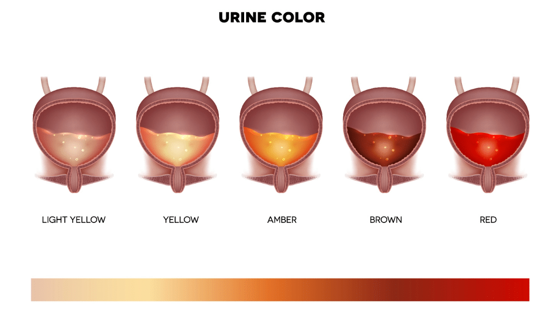
• Urine that is pink, red, or brown

Causes
A pinkish hue in the urine can be caused due to consumption of foods such as beetroot, as well as certain medications, but a change in urine coloration due to these factors is distinct from blood associated with medical problems. The most common medical causes of hematuria include urinary tract infections or bladder infections, viral diseases such as hepatitis, kidney stones, bladder stones, bladder or prostate cancer, kidney infections or disease, injury to the kidneys, and benign prostate hyperplasia (in men only). The condition can also be caused by vigorous exercise or sexual activity. Gross hematuria can be detected visibly if the urine turns pinkish or reddish in color. Microscopic hematuria can only be detected during a lab analysis of the urine. Patients with gross hematuria may also experience blood clots in the urine that can lead to an inability to pass urine, causing pain.
Hematuria Care at Integrative Urology
Treatment
Integrative Urology can determine the cause of hematuria and offer effective treatment. Many times, the cause of hematuria is benign, but it is important to receive a prompt evaluation from a medical professional. When you come in for your consultation at Integrative Urology, we will examine your medical history and ask you questions about your lifestyle and activities to determine the probable cause of hematuria. After this examination, a urinalysis will be performed where your urine sample will be analyzed to determine the cause of blood in your urine. It may be necessary to order a blood test to check if your kidneys are functioning properly and removing body wastes. Apart from urine and blood tests, you may also need to undergo additional tests such as a computed tomography (CT) scan, kidney ultrasound, or cystoscopy (a brief camera test) to diagnose the underlying cause of the presence of blood in the urine.
It is important to undergo a thorough evaluation in cases of microscopic and gross hematuria, as it allows for detection and treatment of serious urologic conditions at their earliest stages.
Cystoscopy
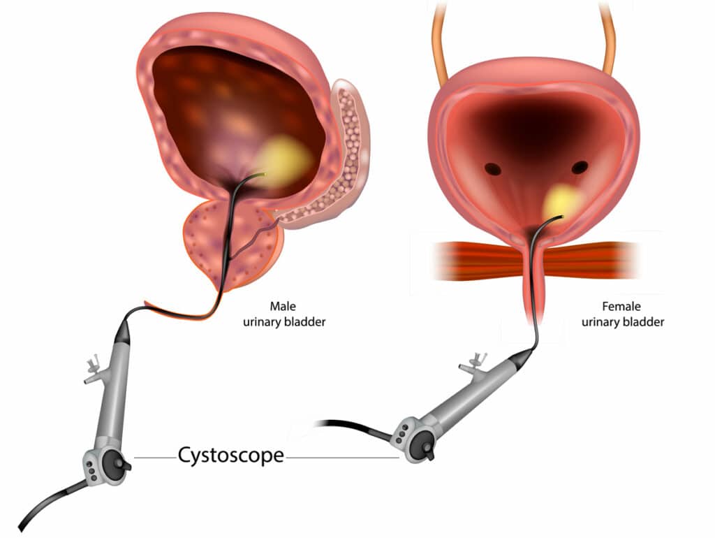
Cystoscopy involves the examination of the inside of the bladder, prostate (in men), and urethra using a cystoscope, a thin, flexible tube with a light and camera on the end. It can provide detailed information about the size, shape, and condition of the bladder, prostate, and urethra, and help guide treatment decisions.
During a cystoscopy, the cystoscope is inserted into the urethra and quickly advanced into the bladder. The camera on the end of the cystoscope transmits images to a monitor, allowing the urologist to examine the bladder, prostate, and urethra for abnormalities such as tumors, stones, prostate enlargement, or infections. It can also be used to collect tissue samples or remove small growths during the procedure for further testing.
Cystoscopy is usually performed in the office under local anesthesia with an option for nitrous oxide analgesia (laughing gas), depending on the patient's preference and the complexity of the procedure. The duration of the procedure can vary depending on the reason for the cystoscopy but typically lasts between 1 to 5 minutes. Most patients tolerate the procedure with mild discomfort, though some may experience significant discomfort. After the procedure, the patient may experience burning during urination, as well as some blood in the urine. These symptoms usually subside within a few hours to a day.

